What is Penile Function Test?
Penile function or impotence test is used for the determination of whether penile function disorders such as Erectile Dysfunction (ED) are of vascular or vasculogenic nature. Several causes may be responsible for Erectile Dysfunction. The main causes of erectile failure include vasculogenic, neurogenic, hormonal, and psychological factors.
How to Perform Penile Function Tests
The purpose of penile physiological tests is to diagnose vascular causes of ED or rule this factor out. All common risk factors for cardiovascular disease also serve as vasculogenic risk factors for impotence or penile function failure.
The physiological assessment of penile function includes the measurements of penile blood pressure and the corresponding penile-brachial index (PBI), Pulse Volume Recordings (PVR) on the penis, Doppler blood flow measurements on the penile arteries, and particularly the penile dorsal arteries, and PPG penile measurements.

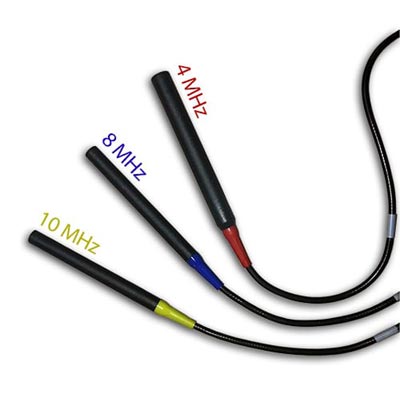
Doppler Probes
10 MHz Probe is the Ideal Choice for this Test

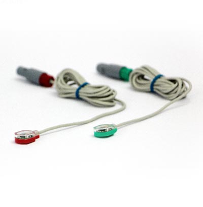
PPG Sensors
Secondary Method of ABI Assessment

Inflatable Cuffs
High quality available in a variety of sizes
Using the Falcon for Penile Function Tests
The Falcon has a dedicated Penile function protocol that allows complete assessment and diagnosis of Erectile Dysfunction and vasculogenic sources of impotence. The Falcon supports all standard methods for penile physiological testing and diagnosis, including measurements of penile pressure and calculation of the corresponding PBI Penile-Brachial Index, PVR, Doppler, and PPG penile measurements. In addition, the Falcon supports Reactive Hyperemia, and all of the physiological tests can then be repeated under RH stress conditions for improved diagnosis.
The Falcon has an assortment of small pressure cuffs of various sizes that are designed for penile pressure and PVR testing. In addition, a range of Doppler probes of various frequencies is available for penile blood flow testing, and in particular, the 10 MHz probe is considered ideal for measurements in superficial arteries. Special small disk PPG sensors can be ideal for blood pressure measurements as well and can be attached to the penis via a special dedicated, transparent adhesive sticker.
The Penile Function protocol can be configured to include a combination of any number of the physiological tests described above for optimal and complete vascular and physiological diagnosis for the vasculogenic sources of impotence. PBI is calculated automatically and clearly displayed both on-screen and in the examination report. All Doppler blood flow velocity parameters are also displayed automatically for each Doppler measurement. These include:
- Peak blood flow velocity,
- Mean blood flow velocity,
- End-Diastolic blood flow velocity,
- Pulsatility Index,
- Resistance Index,
- Systolic to Diastolic Ratio,
- Systolic Rise Time.
For PVR assessment, the Falcon provides the waveform Amplitude and Systolic Rise Time parameters, in addition to the high-resolution PVR waveform.
Expected Results
PBI is defined as the penile blood pressure divided by the higher of the right/left brachial pressure. According to several international guidelines PBI is diagnosed and evaluated as follows:
| Range | Common Diagnosis |
| 0.7 ≤ PBI < 1.0 | Normal |
| 0.6 ≤ PBI < 7 | Borderline |
| PBI ≤ 0.6 | Abnormal |
For Doppler assessment of penile function, a peak systolic blood flow velocity above 30 cm/s is generally considered normal. In addition, an RI (Resistance Index) above 0.8 is also considered normal.
PVR measurements that are considered normal include a short systolic rise time and a high amplitude, with a Dicrotic Notch in the waveform that may be present or absent.
Selected Literature
Guidelines on Male Sexual Dysfunction: Erectile dysfunction and premature ejaculation, E. Wespes et al., European Association of Urology 2012
Techniques in noninvasive vascular diagnosis, An Encyclopedia of Vascular Testing, Robert J. Daigle, Summer Publishing LLC., Third edition Nov 2008, Ch. 14, pp. 239-246
Noninvasive vascular evaluation in male impotence: Technique, Cindy Ramirez, Mike Box and Leonard Gottesman, Bruit, Vol IV, June 1980, pp. 14-16
A Comparison of Penile-Brachial Index (PBI) and Penile Pulse Volume Recordings for Diagnosis of Vasculogenic Impotence, Donna Stauffer and Ralph G. Depalma, Bruit, Vol VII, March 1983, pp. 29-31
The noninvasive diagnosis of vasculogenic impotence, Bruce M. Elliott et al., J VASC SURG 1986; 3:493-7
Usefulness of power Doppler ultrasonography in evaluating erectile dysfunction, A.J. Golubinski and A. Sikorski, BJU International (2002), 89, 779–782

What is Pulse Wave Velocity?
Pulse Wave Velocity (PWV) is a simple and non-invasive measurement, which can be measured at various locations along the arterial circulation to assess arterial stiffness.
Pulse wave velocity (PWV) is widely recognized as a simple and reliable clinical measure of arterial stiffness/elasticity, which is correlated with vascular disease. The contractions of the heart, which drive the arterial blood, also generate arterial blood pressure pulse waves which propagate through the arterial walls. PWV is defined as the velocity at which these arterial blood pressure pulses propagate.
How to Measure Pulse Wave Velocity
While there is no one set standard of how or where to measure PWV, some of the more common PWV measurements include the baPWV, which reflects the pulse wave propagation between the brachial and ankle, cfPWV reflecting propagation between the carotid and femoral, and faPWV also know as leg PWV reflecting the PWV along the leg between the femoral and ankle.
The measurement of PWV is usually a straightforward calculation based on the definition of velocity, i.e., distance divided by time. A physiologic signal, typically the initial systolic upstroke, serves as a time marker. As the arterial pulse wave propagates along the arterial circulation, a short time delay is generated between the proximal and the distal measurement sites. The distance between the 2 measurement sites can be either measured directly or estimated based on some parameters such as patient height. When the PWV is not measured along a straight arterial line, such as the cfPWV, various assumptions need to be made.

Inflatable Cuffs
High quality available in a variety of sizes
Using the Falcon for PWV measurements
The Falcon has a dedicated PWV protocol, allowing a simple and non-invasive diagnosis of various PWV parameters such as the baPWV or faPWV. The measurement is performed with the aid of blood pressure cuffs. These cuff are wrapped around the target measurement sites, such as:
- The Ankle and Thigh for faPWV, or
- Brachial and ankle for baPWV.
The patient is in the supine position during the measurement, and the pressure cuffs are inflated to a venous occlusion pressure, typically around 65 mmHg. The pressure is kept steady while the corresponding PVR waveforms are displayed as a function of time. The examiner can determine the target inflation pressure.
Typically, the examination lasts a few minutes, during which multiple measurements for each cardiac cycle are performed, and the time delay between the 2 measurement cuffs is automatically determined. The distance between the pressure cuffs is entered manually or calculated automatically based on known relationships. Once the examiner stops the measurement process, PWV is calculated automatically and displayed on the screen as an average of the measurements over the approved cardiac cycles.
The Falcon allows to perform the PWV test bilaterally, i.e., simultaneous measurements for the right and left sides. Automatic time markers clearly display the time gap between the proximal and distal systolic rise time to verify the measurement process.

Expected Results
PWV is a quantitative parameter reflecting arterial stiffness or elasticity. Higher PWV values reflect greater than normal arterial stiffness.
This parameter is highly dependent on the method of measurement, as well as on the measurement sites. Therefore, typical values for baPWV differ from faPWV and cfPWV.
In addition, PWV is highly dependent on patient age. Older patients are expected to have more stiff arteries and, as a result, higher PWV values under normal conditions. Diabetic patients are also expected to have a significant increase in PWV values.
Based on literature reports, normal PWV values can range between around 6 m/s to 14 m/s, depending on the method of measurement and the patient’s age.
Example of a Pulse Wave Velocity examination measured using Viasonix Falcon/PRO
Selected Literature
Familial Tendency for Hypertension is Associated with Increased Vascular Stiffness, Wexler et al., Journal of Hypertension 2020, 38.
Peripheral vascular disease assessment in the lower limb: a review of current and emerging non‑invasive diagnostic methods; Shabani Varaki et al, BioMed Eng OnLine (2018) 17:61
Determinants of pulse wave velocity in healthy people and in the presence of cardiovascular risk factors: ‘establishing normal and reference values’, Boutouyrie et al., European Heart Journal (2010) 31, 2338–2350
Brachial–ankle pulse wave velocity: an index of central arterial stiffness?, Sugawara et al., Journal of Human Hypertension;19, 401–406, February 2005
Arterial path length estimation on brachial-ankle pulse wave velocity: validity of height-based formulas, Sugawara et al., Journal of Hypertension 2014, 32:881–889
Arterial stiffness predicts cardiovascular outcome in a low-to moderate cardiovascular risk population: the EDIVA (Estudo de DIstensibilidade VAscular) project, Joao Maldonado et al., Journal of Hypertension 2011, 29:669–675
Effects of aging on leg pulse wave velocity response to single-leg cycling, Sugawara et al., Artery Research (2010) 4, 94e97
Association between estimated pulse wave velocity and the risk of stroke in middle-aged men, Sae Young Jae et al., International Journal of Stroke, 0(0) 1–5, 2020.

What is Extracranial Exam?
Extracranial Doppler examinations are measurements of blood flow velocities in the extracranial vessels, and particularly the Common, External, and Internal Carotid arteries, as well as the Subclavian artery. These measurements are key in the diagnosis of cerebral pathology and circulation.
How to Perform Extracranial Tests
The extracranial vessels are easily accessible with standard Continuous Wave (CW) Doppler probes and allow quick identification of abnormal blood flow patterns. The selection of the Doppler probe frequency depends on the vessel size and its distance from the skin. The 4 MHz and 8 MHz frequencies are the most common selections for extracranial measurements.
Extracranial measurements serve various clinical purposes, such as identifying carotid stenosis or similar lesion, determining increased distal cerebral resistance, Subclavian Steal Syndrome, and Stroke assessment. The measured blood flow velocities increase significantly in the area of a stenosis, with a greater increase as the degree of stenosis increases. This continues until the stenosis reaches a critical level, after which blood flow velocities will decrease as overall blood flow through the artery is compromised. Blood flow velocities will drop finally to zero with a complete 100% occlusion.
Another common parameter is the Lindegaard Ratio (LR). The LR is defined as the ratio between the mean Middle Cerebral artery (MCA) velocity and the mean Internal Carotid Artery (ICA) velocity. It is an essential diagnostic parameter when determining if high cerebral velocities result from a cerebral hemorrhage, arterio-venous malformations, vasospasm or other factors.

Doppler Probes
Gold Standard ABI Measurement Method
Using the Falcon for Extracranial Measurements
The Falcon supports a variety of CW Doppler probes, including 4 MHz, 8 MHz, and 10 MHz probes.
These frequencies allow optimal access and measurements of larger/smaller and deeper/superficial blood vessels. The higher frequencies are intended for smaller and shallower vessels, and vice versa.
The Falcon protocols allow the configuration of any blood vessel of interest, and this is supported with a schematic picture of the carotid circulation for improved documentation.
The Doppler measurements include complete spectral analysis, which allows optimal diagnosis and visualization, for example, of systolic bruits. A complete set of Doppler parameters is automatically calculated, including:
- Peak velocity,
- Mean velocity,
- Diastolic velocity,
- Pulsatility index (PI),
- Resistance index (RI),
- Systolic-Diastolic ratio (S/D),
- Systolic rise time, and
- Heart rate.
The spectral color palette, display, and noise rejection can be configured and optimized for each user.
Likewise, the peak systolic envelope can be controlled, Y-Axis units (cm/sec or KHz), as well as a range of other Doppler controls, including Sweep Time, Gain, High-Pass Filter, Scale, and Volume.

Expected Results
The criteria for the assessment of the extracranial blood flow measurements depends on the pathology. Focal stenosis will cause a significant increase in mean and peak blood flow velocities, up to a critical level, after which flow will decrease. An intracranial hemorrhage and increased MCA velocity will likely result in higher Lindegaard ratio values. Other criteria for the assessment of pathology are further defined in the accepted international guidelines.
Example of Extracranial measurements performed with Viasonix Falcon/PRO on the Common Carotid.
Selected Literature
Cerebrovascular Ultrasound in Stroke Prevention and Treatment, Edited by Andrei V. Alexandrov, Blackwell Publishing 2004
Ultrasound Assessment of the Intracranial Arteries, Darius G. Nabavi et al., in “Introduction to Vascular Ultrasonography”, Ed. Pellerito and Polak, Elsevier Health Sciences, 2012, Ch 12, pp 202-228
Transcranial Doppler, Peter J. Kirkpatrick and Kwan-Hon Chan, Head Injury. Edited by Peter Reilly and Ross Bullock. Published in 1997 by Chapman & Hall, Ch 13, pp 243-259


