- Cerebral Monitoring
- Emboli Detection
- Endarterectomy
- Evaluation of ICP changes
- Intraoperative Doppler
- Intraoperative TCD Monitoring

Cerebral Monitoring
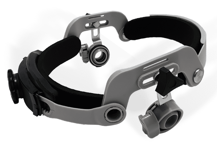
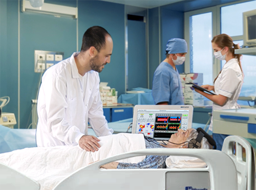
Cerebral monitoring using 2 MHz Transcranial Doppler sonography is the short- or long-term TCD monitoring of blood flow in one of the blood vessels comprising the Circle of Willis, typically the MCA (Middle Cerebral Artery).
TCD Monitoring is an important tool for detecting cerebral ischemia as well as following symptomatic carotid stenosis or monitoring during Carotid Endarterectomy, coronary artery bypass graft surgery (CABG), and other surgeries that require anesthesia. Using transcranial doppler is well recognized by many clinical international societies, including the American Heart Association (AHA).
Cerebral monitoring can be either unilateral or bilateral.
Unilateral monitoring follows the changes in the blood flow velocity on one side of the brain.
Bilateral monitoring is the simultaneous measurement of flow on both the right and left sides of the brain, typically the right and left middle cerebral arteries.
During Transcranial Doppler ultrasonography monitoring in the middle cerebral artery, the focus is on following the trends in velocities over time, timely detection of changes in Intracranial Pressure (ICP), or identifying special events during the monitoring session such as embolic events.

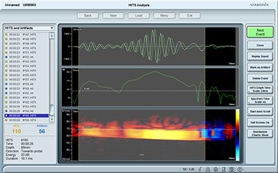
Emboli Detection
Cerebral emboli are particles or air bubbles that originate anywhere in the human body and travel through the arterial circulation into the arteries of the brain.
These particles or air bubbles may clot and occlude the brain’s arteries, resulting in a sharp local decrease in brain blood flow that can cause a stroke. Cerebral emboli can be either solid or gaseous:
- Solid emboli, such as atherosclerotic plaques or blood clots, typically originate in the carotid arteries or from the heart.
- Air bubbles (gaseous emboli) can originate from sources outside the body, such as during surgical procedures or internally, for example, from mechanical Aortic valves or during decompression.
Endarterectomy
Carotid Endarterectomy (CEA) is a surgical procedure designed to remove plaque in the Carotid artery by separating it from the carotid arterial wall. Thus, carotid arterial constriction, which may cause a decrease in cerebral perfusion as well as a potential source for cerebral emboli, is removed.
During a CEA procedure, Transcranial Doppler (TCD) monitoring plays a crucial role in ensuring optimal cerebral perfusion and minimising potential risks, such as the early detection of atherosclerotic or thombotic emboli that may be dislodged during surgery and travel into the brain.
Evaluation of ICP changes
Intracranial Pressure (ICP) is the pressure inside the skull, which has a direct effect on the blood perfusion of the brain.
Cerebral Perfusion Pressure (CPP) is a function of the difference between the Mean Arterial Pressure (MAP), which is the driving pressure, and the opposing pressure, which is the ICP.
Optimal CPP is defined as a pressure ≥70 mmHg [2].

Intraoperative Doppler
Intraoperative Doppler measurements are the detection of blood flow velocity in small micro-vessels inside the sterile field during surgery. Typically a micro-Doppler probe with a frequency of 16 MHz or 20 MHz is used.
The probe is placed directly on the wet tissue or the external wall of the blood vessel.

Intraoperative TCD Monitoring
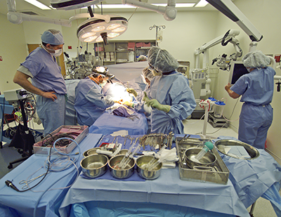
Intraoperative monitoring with Transcranial Doppler (TCD) equipment refers to continuous measurements of cerebral blood flow velocities and pulsatility indices during surgical procedures.
Intraoperative TCD measurements are designed to ensure proper blood supply to the brain throughout the surgery, count the number of emboli that pass to the brain and monitor various specific procedures during the surgical process.
For these reasons, both anesthesiologists and surgeons are particularly interested in Intraoperative monitoring.


